Congenital Scoliosis
What is congenital scoliosis?
The word congenital means scoliosis appearing on a newborn child. Congenital scoliosis occurs due to problems based on the development of vertebrae in unborn babies (between 4th-8th weeks). Due to incomplete formation or a failure of separation of one or more vertebrae cause congenital scoliosis. There may be accompanying abnormalities in the spinal cord (41%), heart (7-12%) or kidneys (20%).
What is the frequency of congenital scoliosis?
Scoliosis is classified according to the cause of occurrence. The most common type of scoliosis is called idiopathic scoliosis. Second most common is congenital scoliosis. Its frequency is 1-4% in the society.
What causes congenital scoliosis?
Among the causes of congenital scoliosis, we may point out:
- Infections in the mother’s womb
- Vitamin & mineral deficiencies
or the mother’s
- Diabetes
- Cardiac diseases
- Hyperthermia
- Use of alcohol
- Valproic acid poisoning
Congenital scoliosis can also be an accompanying disease to other congenital diseases.
What are structural defects that cause congenital scoliosis?
Structural defects can cause scoliosis in several ways. The vertebrae are like short cylinders aligned on top of one another. A pyramid-like half vertebra between other vertebrae causes the spine to bend on this point and form scoliosis. It can also emerge when some adjacent vertebrae grow attached to each other while other (separated) vertebrae are growing much bigger. Sometimes, the problems in the ribcage (ribs being attached to each other) can cause asymmetrical growth of the vertebral column, leading to congenital scoliosis as well.
How is the process of congenital scoliosis?
Congenital scoliosis is generally progressive. So it is important to have an early diagnosis. Parents need to consult a doctor right away if they notice a difference in the baby’s neck, back or waist. Congenital scoliosis treatment in early ages may require surgical intervention.
Children that have congenital scoliosis may also develop a secondary curvature above or under the primary curvature in order to secure physical integrity and balance. This curvature may become bigger than the primary curvature in time. Monitoring curvatures is very important for the therapy.
What should be the monitoring intervals in congenital scoliosis?
The problem in the vertebra, the location of the problem, the patient’s age are very important in deciding the treatment method for congenital scoliosis. The spine grows very fast between the ages of 0-5 and 10-15. These periods require a much more frequent monitoring of congenital scoliosis. The interval in these periods should be 6 months maximum. The treatment method should be decided according to the increase in deformity.
What are symptoms for congenital scoliosis?
- Hirsutism (Increased hair development) in the lower back
- Asymmetrical skin tags in the back
- Different color of the skin
- Asymmetrical alignment of the vertebrae in posterior view
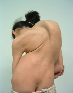
- One side hump in the back when the patient bends forward
- Abnormal palpable bone spurs in the back
Which tests are made for congenital scoliosis patients?
X-rays films are generally requested after a detailed physical examination. We can view the whole vertebral column in this radiograph which is also called scoliosis graphy (imaging). Computerized tomography (CT-Scan) and magnetic resonance (MR) imaging are also required. The films allow the measurement of scoliosis angle.
The main elements that will determine the treatment method of scoliosis are the curvature’s angle and location, the patient’s age, the existence of an accompanying kyphosis or lordosis. Patients who are diagnosed with congenital scoliosis based on these parameters can be monitored periodically with the help of radiographic imaging or they can undergo surgical treatment.
What is the purpose in congenital scoliosis treatment?
- Correcting spine deformities
- Preventing further spine deformities
- Protecting the spine’s growing potential and letting it grow
- Providing the development of ribcage
- Protecting the functionality of lungs and heart
- Preventing short stature and other cosmetic or psychological problems
What is done during the monitoring of congenital scoliosis?
The patients of congenital scoliosis are monitored at 6-month intervals if the curvature in their spine is small and not progressive. Detecting an increase during the monitoring can provide a timely transfer of the patient to surgical treatment. Sometimes a curvature in the spine based on a half vertebra is supported by another half vertebra in the opposing side. This situation is considered as balancing and only requires to be examined at certain intervals and with x-ray imaging.
A congenitally deformed spine may not always cause a curvature. Even an experienced surgeon cannot tell if the congenital scoliosis is likely to progress or not. Therefore congenital scoliosis must be monitored. The angle of scoliosis is measured by x-rays at the follow-up examinations and compared with previous measurements. If there is a progress in congenital scoliosis, it is thereby noticed and early surgical intervention becomes possible.
What are the treatment methods for congenital scoliosis?
Brace treatment could be beneficial if the congenital scoliosis has a long curvature which encompasses a large section of the spine. Brace treatment is also beneficial for the treatment of secondary curvatures. But brace treatment is generally unsuccessful because of the curvature being short and inflexible in congenital scoliosis.
Surgical treatment is the only option in progressive cases. It is the most applied method of treatment since most of the cases do progress and brace treatment usually fails.
 What are the types of surgical treatment in congenital scoliosis?
- Hemivertebra resection
Some patients with congenital scoliosis need early surgical intervention. If the scoliosis is caused by an incomplete hemivertebra, this might be resected. The early resection of the hemivertebra that causes congenital scoliosis prevents a curvature from occurring.
- Controlling the curvature by growing rods
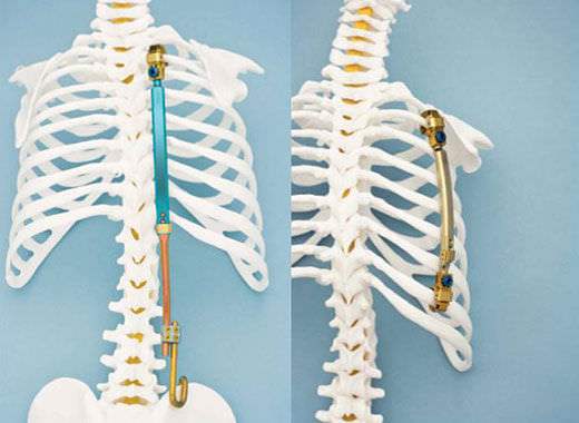
In children with congenital scoliosis, spinal fusion may lead to short stature, or a defective development of lungs and ribs. So it is not considered. Instead, the correction is achieved by the help of growing rods that are placed between the upper and lower ends of the scoliosis curvature. So a spinal fusion becomes unnecessary.
When these rods are implanted, they are lengthened at regular intervals in order to correct the scoliosis. There are two types of these rods (surgically growing and magnetically growing). Patients that use surgically growing rods need to undergo a surgery in every 6 months. This surgical procedure under general anesthesia may lead to some other accessory problems. Therefore magnetically (remotely) growing rods are more popular. These lengthening processes control and correct the curvature until the adulthood, allow the ribs and lungs to grow normally and prevent the patient’s body from short stature. This modern technology (remotely lengthening magnetic rods) can be considered as revolutionary especially for little aged patients of scoliosis since it makes the repeating surgeries unnecessary.
- Spinal fusion
The treatment of congenital scoliosis diagnosed in early ages can be different from that diagnosed in adulthood. In scoliosis surgery, the purpose is to correct the curvature and to normalize the spine. The vertebrae are fused together in order to maintain the correction. Spinal fusion in children who have yet an incomplete spine leads to a short stature, compression of the spinal cord and the nerves due to lack of space, defective development of lungs and the heart due to reduced space in the rib cage. Spinal fusion therefore is more suitable for young patients who have completed the development of their spine or adult patients.
- VEPTR (Vertical Expendable Prosthetic Titanium Rib) Surgery
The development of rib cage is very important for patients who have scoliosis and rib abnormalities at the same time, in order to prevent lung failures. Rib cage expansion (VEPTR) is a surgical procedure practiced to correct the scoliosis and to expand the rib cage in adolescence. When sufficient volume for the rib cage is achieved, a spinal fusion surgery is performed at the end of adolescence.


